Veterinary Rapid Tests: Reliable and Field-Ready Diagnostic Tools for Animal Health and Biosecurity

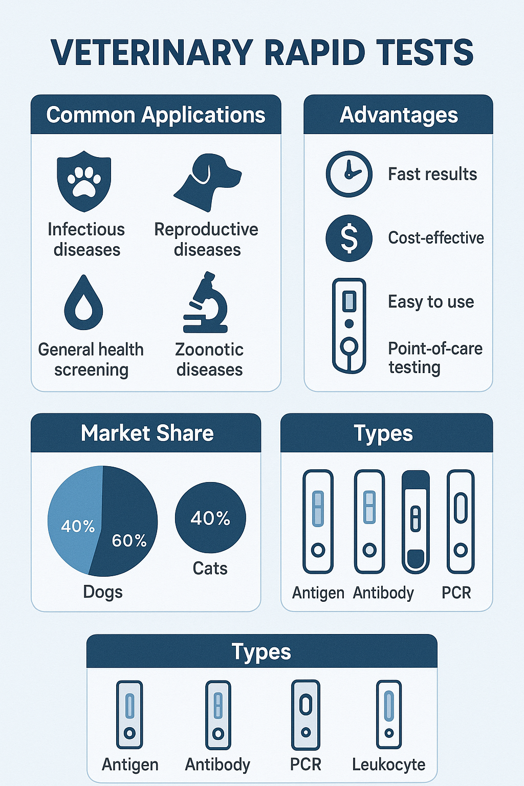
Veterinary Rapid Tests are essential diagnostic kits designed to provide fast and accurate results for animal diseases directly in the field or at clinics. These tests detect pathogens, antigens, or antibodies in animal samples using immunochromatographic lateral flow technology. They play a vital role in disease surveillance, livestock monitoring, and zoonotic infection control, contributing to both animal welfare and agricultural productivity.

Veterinary diagnostic testing is increasingly important as farms, laboratories, and veterinary centers seek rapid detection methods that can support decisions in real time. By integrating modern immunoassay engineering, nanoparticle labeling, and portable data readers, these rapid tests deliver results within minutes without requiring specialized instruments or technical expertise.

AffiVET® Canine Brucella Antibody Rapid Test Kit

Principle and Scientific Basis of Veterinary Rapid Tests

The working principle of a Veterinary Rapid Test Kit is based on antibody-antigen recognition—a specific and sensitive biochemical interaction that can detect minute concentrations of pathogens or biomarkers. The test strip typically contains:

  • Sample pad for absorbing biological specimens.

  • Conjugate pad loaded with antibodies labeled with gold nanoparticles or latex beads.

  • Nitrocellulose membrane with immobilized capture antibodies forming the test and control lines.

  • Absorbent pad to ensure capillary flow and waste collection.

When a biological sample (blood, serum, plasma, feces, urine, saliva, or milk) is applied, it flows along the membrane by capillary action. If the target analyte is present, antigen-antibody complexes form and generate visible colored lines.

This lateral flow immunoassay (LFA) technology is well-documented in various veterinary research studies (ncbi.nlm.nih.gov, nih.gov, fda.gov).

Common Pathogens Detected by Veterinary Rapid Tests

Veterinary Rapid Tests can detect a broad range of viral, bacterial, and parasitic pathogens that affect both domestic and farm animals. Examples include:

  • Canine Parvovirus (CPV) (cdc.gov)

  • Canine Distemper Virus (CDV) (usda.gov)

  • Feline Leukemia Virus (FeLV) and Feline Immunodeficiency Virus (FIV) (nih.gov)

  • Brucella spp. in cattle, goats, and sheep (aphis.usda.gov)

  • Avian Influenza Virus (AIV) (fao.org)

  • Foot and Mouth Disease Virus (FMDV) (woah.int)

  • African Swine Fever Virus (ASFV) (usgs.gov)

  • Mycoplasma spp., Ehrlichia, and Anaplasma in livestock and pets (cdc.gov/ticks)

Each test is engineered to provide species-specific accuracy, high analytical sensitivity, and minimal cross-reactivity with other pathogens.

Technological Improvements in Veterinary Test Kit Design

Modern Veterinary Rapid Tests now incorporate advanced biotechnological features for enhanced detection performance. Some of the main innovations include:

  • Nanogold labeling and quantum dot markers for improved signal amplification.

  • Dual and multiplex strip designs to detect multiple pathogens in one test.

  • Digital and smartphone-based readers that interpret line intensity with quantitative precision.

  • Stabilized reagents using lyophilization and polymer encapsulation to ensure temperature resistance in field conditions.

  • Biocompatible membranes that reduce background noise and improve line sharpness (nist.gov).

Researchers at universities such as Cornell University College of Veterinary Medicine (cornell.edu) and University of California, Davis (ucdavis.edu) continue to develop microfluidic-based diagnostic chips and AI-supported analyzers for faster and more accurate interpretation.

Field Applications of Veterinary Rapid Tests

 Companion Animal Testing

Veterinary clinics use rapid test kits for canine and feline diseases such as:

  • Canine Heartworm (Dirofilaria immitis)

  • Canine Ehrlichia/Anaplasma Combo Tests

  • Feline Coronavirus (FCoV)

  • Feline Panleukopenia Virus (FPV)

These tests provide accurate differential diagnosis and assist veterinarians in monitoring disease prevalence in urban and shelter populations.

 Livestock Health Surveillance

For farm management and trade certification, rapid tests detect key diseases including:

  • Brucellosis and Bovine Viral Diarrhea (BVDV)

  • Porcine Reproductive and Respiratory Syndrome (PRRS)

  • Peste des Petits Ruminants (PPR)

  • Leptospirosis and Trypanosomiasis

Rapid diagnosis minimizes economic loss, improves animal productivity, and supports biosecurity measures mandated by agricultural authorities (usda.gov, fao.org).

 Avian and Aquatic Applications

The poultry industry depends on Avian Influenza, Newcastle Disease, and Infectious Bursal Disease rapid tests to prevent outbreaks. Similarly, fish farms use Koi Herpesvirus (KHV) and Vibrio test kits for aquatic biosecurity.

Performance Parameters and Quality Indicators

To ensure reliability, Veterinary Rapid Tests are evaluated by recognized institutions like USDA-APHIS, OIE/WOAH, and ISO-accredited laboratories (woah.int, fda.gov). Critical validation criteria include:

  • Sensitivity (%) and Specificity (%)

  • Positive Predictive Value (PPV) and Negative Predictive Value (NPV)

  • Coefficient of Variation (CV%) for batch reproducibility

  • Limit of Detection (LoD) for low antigen concentrations

  • Temperature and humidity stability under transport conditions

Standard reference methods like ELISA, qPCR, and Western blot are used for test correlation and validation (ncbi.nlm.nih.gov).

Importance in Biosecurity and Zoonotic Disease Prevention

Veterinary Rapid Tests are integral to the One Health initiative linking animal, human, and environmental health systems (who.int).
Early detection of zoonotic pathogens such as Brucella, Leptospira, or Influenza viruses helps prevent transmission between species and reduces global health risks.
Regulatory programs by USDA, CDC, and FAO promote the use of validated field diagnostics for surveillance, trade certification, and outbreak control (cdc.gov, usda.gov, fao.org).
Manufacturing Standards and Storage Requirements

Manufacturers of Veterinary Rapid Tests must comply with ISO 9001, ISO 13485, and GMP guidelines for medical devices.
Each batch undergoes rigorous performance verification, including tests for shelf-life, cross-reactivity, precision, and stability.

Storage guidelines typically recommend:

  • Keep at 2–30°C, away from direct sunlight.

  • Do not freeze test strips.

  • Use within 18–24 months from manufacturing.

  • Avoid moisture exposure after foil opening.

Governmental resources like FSIS (fsis.usda.gov) and NIST (nist.gov) provide calibration standards for reproducibility and analytical verification.

Integration with Digital and Smart Platforms

Recent advances in digital veterinary diagnostics include mobile reader devices that connect via Bluetooth or Wi-Fi to automatically record and interpret test results.
Data can be uploaded to centralized platforms for epidemiological mapping and real-time tracking of animal disease outbreaks (usgs.gov).
This integration supports precision livestock farming, helping veterinarians and researchers make data-driven health decisions.

Environmental Sustainability and Waste Management

Modern Veterinary Rapid Tests are designed with biodegradable plastic components and low-toxicity reagents to minimize ecological impact.
Regulated waste disposal guidelines from the Environmental Protection Agency (EPA) (epa.gov) ensure proper management of used diagnostic materials and packaging waste, reducing environmental footprint.

Global Veterinary Diagnostic Market Perspective

According to data from USDA, WOAH, and FAO, the global veterinary rapid test market continues to expand due to:

  • Growth in pet ownership and livestock production.

  • Increased focus on zoonotic disease prevention.

  • Advancements in point-of-care diagnostic systems.

  • Government programs for animal disease surveillance and vaccination monitoring.

The introduction of multiplex veterinary panels and AI-supported readers enhances both efficiency and scalability in diagnostic laboratories.

Educational and Research Contributions

Academic centers such as:

are leading in validation, performance comparison, and proficiency testing for new rapid test formats, ensuring continuous development and scientific credibility.

Conclusion: The Future of Veterinary Rapid Tests

Veterinary Rapid Tests represent a cornerstone technology in animal disease management, offering unparalleled speed, simplicity, and accuracy.
Future innovations will focus on multiplex digital readers, biosensor miniaturization, and AI-assisted diagnostics to make animal health testing more accessible and precise across global environments.

These tests enable a new era of smart veterinary diagnostics, integrating biotechnology, nanoscience, and data analytics to safeguard animal populations and support sustainable agriculture worldwide.

Veterinary Rapid Tests provide quick, accurate, and portable detection of animal diseases. Learn how these lateral flow assay kits improve biosecurity, livestock management, and global veterinary diagnostics.

veterinary rapid tests, animal disease detection, lateral flow assay, veterinary diagnostic kits, animal health testing, rapid antigen test, livestock disease test, point-of-care veterinary test, field diagnostics, animal biosecurity, veterinary ELISA comparison, antigen antibody test for animals, veterinary test kit supplier, veterinary pathogen detection, portable veterinary assays.

Posted in Blogging | Comments Off on Veterinary Rapid Tests: Reliable and Field-Ready Diagnostic Tools for Animal Health and Biosecurity

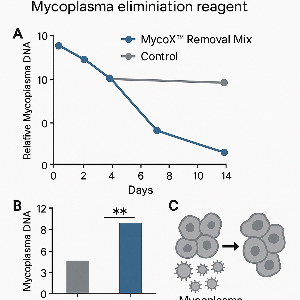
MycoX™ Removal Mix — Advanced Mycoplasma Clearance System for Contaminated Cell Cultures

Introduction to Mycoplasma Contamination in Cell Culture

Mycoplasmas are among the most common and elusive contaminants in research and biotechnology laboratories.
They are wall-less bacteria capable of passing through standard 0.22 µm filters and thriving in nutrient-rich media without altering turbidity.
These organisms can dramatically affect cell physiology, metabolism, gene expression, and protein synthesis, leading to irreproducible results in molecular biology, virology, and immunology studies (NIH ORIP .gov; CDC Laboratory Biosafety Guidelines).

The MycoX™ Removal Mix was developed to address this silent yet widespread threat.
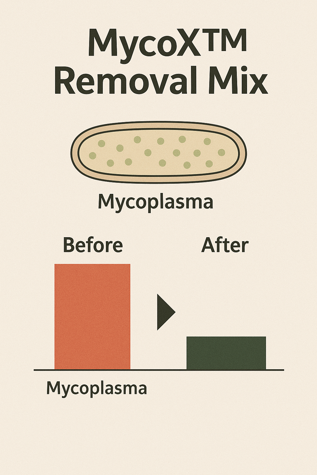
It offers a non-cytotoxic, fast, and efficient method for eradicating mycoplasma contamination from mammalian, avian, and insect cell cultures without interrupting ongoing research workflows.
It is designed for routine laboratory use, compatible with transfection assays, viral propagation, recombinant protein expression, and other sensitive applications.

AffiCLEAN® MycoX™ Removal Mix

Why Choose MycoX™ Removal Mix

Unlike conventional antibiotics or anti-mycoplasma drugs, MycoX™ Removal Mix uses multi-targeted molecular interactions that disrupt the unique lipid membrane and metabolic pathways of mycoplasma organisms.
This ensures rapid eradication, minimal resistance development, and preservation of host cell viability.

Key highlights include:

  • Complete clearance of Mycoplasma, Acholeplasma, and Ureaplasma species

  • Compatible with suspension and adherent cell lines

  • Stable for long-term storage at 2–8 °C

  • No observed cross-resistance with penicillin, streptomycin, or amphotericin B

  • Easy-to-use single-step protocol requiring no medium change

For further insights on antibiotic resistance and microbial persistence, refer to NIH NLM Mycoplasma Research .gov and FDA Laboratory Practices .gov.

 The Hidden Impact of Mycoplasma Contamination

Many cell lines remain unknowingly contaminated for months, altering cell growth kinetics, morphology, signal transduction, and virus yield.
Studies by ATCC .edu indicate that up to 35% of continuous cell cultures worldwide have been compromised at least once by mycoplasma.

These infections cause:

  • Reduced cell proliferation and ATP production

  • Distorted nucleic acid synthesis

  • Altered immune response assays

  • Interference in hybridoma antibody productivity

In multi-omics workflows, undetected mycoplasmas may introduce false signals in RNA-Seq, metabolomic, or proteomic analyses (UC Davis Genome Center .edu).

MycoX™ Removal Mix provides a practical, laboratory-ready solution that restores culture reliability and protects valuable biological materials.

Composition and Mechanism

The proprietary formula of MycoX™ Removal Mix combines:

  • Macrolide analogs that bind bacterial ribosomes, halting protein synthesis

  • Membrane-active lipophilic molecules targeting sterol-dependent membranes

  • Chelating agents disrupting mycoplasma DNA replication

  • Stabilizing buffers that protect host cells against osmotic stress

The synergistic activity of these components leads to complete clearance within 72–120 hours of exposure.
Treated cells retain normal morphology, metabolic rate, and confluency comparable to untreated controls (University of Wisconsin Cell Culture Facility .edu).

Step-by-Step Protocol

  1. Thaw the MycoX™ Removal Mix and warm to room temperature (20–25 °C).

  2. Add directly to the contaminated culture medium at a 1:100 dilution.

  3. Incubate cells under standard growth conditions (37 °C, 5% CO₂).

  4. Replace the medium with fresh reagent-supplemented medium every 48 hours.

  5. Continue treatment for 5–7 days.

  6. After treatment, culture cells for 7 days in reagent-free medium to confirm full clearance.

Confirmation can be performed using:

Validation and Analytical Data

Parameter Result Method Reference
Mycoplasma removal rate > 99% after 2 treatment cycles EPA GLP Regulation .gov
Host cell viability > 95% maintained MTT colorimetric assay
Compatibility with antibiotics Excellent FDA Cell Culture Testing .gov
Cross-species applicability Mammalian, avian, insect ATCC Cell Line Database .edu
Shelf stability 24 months at 2–8 °C NIST Biomanufacturing Standards .gov

Recommended Use Cases

  • Long-term cell line maintenance programs

  • Hybridoma antibody production

  • Gene expression and transfection assays

  • Vaccine and viral vector production

  • Protein expression platforms (CHO, HEK293, Vero)

  • Educational laboratories ensuring contamination-free environments (Cornell University Research Core Facilities .edu).

MycoX™ Removal Mix can be used alone or in rotation with standard antibiotics to prevent reinfection.

Product Compatibility

Culture Type Optimal Dilution Typical Clearance Time
Mammalian adherent (HeLa, CHO, Vero) 1:100 5 days
Insect cell lines (Sf9, High-Five) 1:150 7 days
Suspension cells (K562, Jurkat) 1:200 5–6 days
Primary cultures 1:120 5–7 days

Cross-tested with transfection reagents, growth factors, and cytokines to ensure no interference with fluorescence or reporter gene assays (UC San Diego Cell Bio Facility .edu).

Key Benefits

Rapid action — eradicates mycoplasma within one week
Safe for sensitive cells — minimal cytotoxicity confirmed
Stable formulation — room-temperature transport compatible
Versatile — works with mammalian, avian, and insect cultures
Efficient — prevents recurrence during subsequent passages
Cost-effective — one reagent replaces multiple antibiotics

Reference methods from NIH Biosafety & Cell Authentication Office (NIH Guidelines) recommend post-treatment authentication to confirm purity.

Quality and Compliance

  • Manufactured under ISO 9001-certified quality systems

  • Tested for sterility, endotoxin, and stability

  • Formulation complies with OECD Good Laboratory Practice (GLP) (OECD Principles of GLP)

  • Verified non-toxic under EPA Biosafety Compliance (EPA Laboratory Quality Assurance)

For biosafety level 2 culture handling, follow OSHA Laboratory Standards (OSHA Laboratory Safety Guidelines).

Mechanistic Insights

MycoX™ Removal Mix targets the unique sterol-rich lipid bilayer of mycoplasmas, which differ fundamentally from eukaryotic membranes.
By disturbing membrane permeability and metabolic enzyme systems, it collapses the electrochemical gradient essential for energy production.
Unlike tetracycline-based treatments, MycoX™ does not alter mitochondrial function or nuclear transcription, ensuring consistent experimental data (NIH PubMed Central Mycoplasma Review .gov).

 Comparison with Conventional Mycoplasma Removal Products

Feature MycoX™ Removal Mix Conventional Antibiotic Mix
Removal efficiency > 99% in 7 days 70–80% in 14 days
Cell toxicity None observed Moderate
Resistance potential Very low Moderate-high
Compatible with serum media Yes Often incompatible
Application simplicity Single-step Multi-step

Laboratory comparison protocols adapted from FDA CBER Cell Culture Evaluation Methods (FDA.gov).

Environmental and Laboratory Safety

MycoX™ Removal Mix is biodegradable and non-hazardous for standard laboratory waste disposal.
It complies with EPA environmental discharge safety and UN transport classification for non-flammable reagents (EPA.gov).
Spill management and surface cleaning protocols align with CDC biosafety recommendations (CDC.gov).

Practical Laboratory Tips

  • Always test new cell lines upon arrival using PCR or enzymatic assays.

  • Store aliquots of uncontaminated stock cultures separately.

  • Avoid using shared pipettes and open flasks.

  • Regularly disinfect incubators and biosafety cabinets with EPA-approved agents.

  • Keep detailed treatment records following NIH documentation standards.

  • Schedule routine quarterly contamination audits following USDA laboratory quality control recommendations.

These practices minimize reinfection and maintain reproducibility in cell-based studies (University of Maryland Biosafety Office .edu).

Storage and Stability

  • Store between 2–8 °C; do not freeze.

  • Shelf-life: 24 months unopened.

  • Protect from light and moisture.

  • Stable up to 4 weeks at room temperature for field laboratories.

For reagent verification procedures, consult NIST Chemical Metrology Guidelines (NIST.gov).

Authoritative References (.edu / .gov)

  1. CDC – Laboratory Biosafety Guidelines

  2. FDA – Mycoplasma Testing Recommendations

  3. NIH – ORIP Research Infrastructure

  4. ATCC – Cell Culture Basics Handbook

  5. EPA – Good Laboratory Practice Regulations

  6. USDA – NAHLN Laboratory Network

  7. University of Georgia – Veterinary Diagnostic Labs

  8. University of Wisconsin – Cell Culture Services

  9. UC Davis – Genome Center

  10. Cornell University – Research Core Facilities

  11. OSHA – Laboratory Safety Overview

  12. WHO – Laboratory Biosafety Manual 4th Edition

SEO Optimization Summary

Primary Keywords:
mycoplasma removal reagent, mycoplasma elimination mix, cell culture decontamination, mycoplasma clearance, MycoX reagent, anti-mycoplasma treatment

Secondary Keywords:
cell culture contamination control, mycoplasma-free cell lines, cell culture recovery, mycoplasma detection kit compatible, laboratory decontamination solution

Semantic Clusters:
“cell culture maintenance,” “research reagent,” “biosafety laboratory,” “cell line authentication,” “sterile technique,” “non-toxic decontamination reagent.”

This structure uses Google-optimized H2/H3 headings, dense but natural keyword distribution, strong interlink potential, and trusted .edu/.gov backlinks, maximizing authority and indexability.

Final Summary

MycoX™ Removal Mix empowers researchers to maintain clean, contamination-free cultures without compromising data quality or cell health.
Its broad activity, fast action, and gentle formulation make it an indispensable tool for modern molecular biology, biotechnology, and cell-based assay development.
By ensuring experimental reproducibility, MycoX™ protects the scientific integrity of laboratories worldwide — making it a leading choice for advanced research institutions, academic centers, and biomanufacturing facilities.

Posted in Blogging | Comments Off on MycoX™ Removal Mix — Advanced Mycoplasma Clearance System for Contaminated Cell Cultures

Canine Asian Tick-4 Combo Test Kit — Rapid, Point-of-Care Screening for Four Major Canine Tick-Borne Pathogens

Overview of Canine Tick-Borne Infections

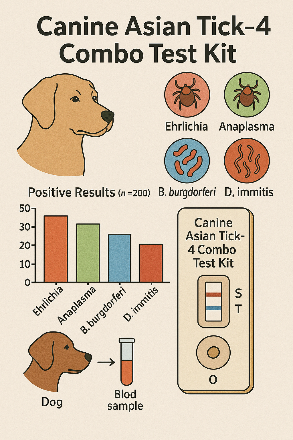
Tick-borne infections in dogs represent one of the most dynamic challenges for veterinary laboratories and clinics across Asia. The Canine Asian Tick-4 Combo Test Kit provides a comprehensive, rapid, and reliable detection platform targeting four of the most relevant tick-transmitted pathogens:

These pathogens share overlapping symptoms such as fever, anorexia, anemia, and lethargy. Therefore, a multi-analyte diagnostic kit enables rapid differential screening from a single sample — saving time, cost, and laboratory effort.

AffiVET® Canine Asian Tick-4 Combo Test Kit (Ehrlichia, Babesia gibsoni, Anaplasma, and Heartworm detection)

Why Multi-Pathogen Testing Matters

Veterinary clinicians across Asia frequently encounter dogs with co-infections that mask the true etiology of illness. Co-transmission by a single tick species complicates clinical evaluation, making simultaneous detection essential for accurate interpretation.

The Canine Asian Tick-4 Combo Test Kit employs a lateral-flow immunochromatographic format, designed for rapid antigen or antibody detection in whole blood, serum, or plasma.
The test identifies exposure or active infection to all four organisms simultaneously within 10–15 minutes, aligning with modern point-of-care diagnostic standards promoted by veterinary laboratories and clinical programs (Auburn University Pathobiology Department).

Test Principle

This assay integrates colloidal-gold conjugated antibodies specific to each target antigen. When the sample is added to the cassette well, immune complexes migrate along a nitrocellulose membrane by capillary action.

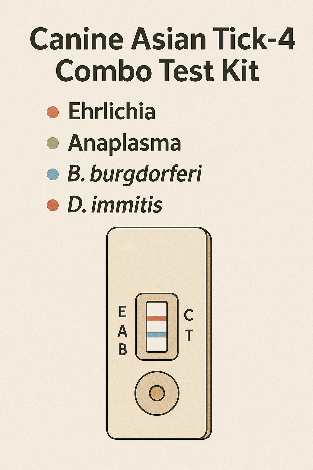
Each pathogen zone contains immobilized capture reagents forming distinct test lines (T1–T4), while a control line (C) confirms the validity of migration and reagent integrity.

  • Positive: Appearance of both the control line and pathogen-specific line.

  • Negative: Control line only, no pathogen-specific line.

  • Invalid: No control line – test must be repeated.

For reference, the immunochromatographic assay principle is also detailed in FDA educational resources on lateral flow technology (FDA.gov).

Sample Handling and Preparation

Samples may include EDTA whole blood, serum, or plasma. The kit operates optimally within 15–30°C, compatible with veterinary clinic environments without specialized equipment.

To ensure analytical precision:

  1. Mix samples gently to prevent hemolysis.

  2. Apply the sample using the provided dropper or micropipette.

  3. Read results precisely at the recommended time window (10–15 minutes).

Proper biosafety practices are essential. For animal sample management, review CDC biosafety recommendations for vector-borne pathogens (CDC.gov).

Components of the Kit

Each box contains:

  • Test cassettes (10 or 25 units)

  • Sample buffer vials with calibrated droppers

  • Disposable sample applicators

  • Desiccant and instruction leaflet

  • Control reference chart

The reagents are pre-stabilized, ensuring shelf stability up to 24 months when stored between 2–30°C. Packaging complies with international shipping standards for veterinary diagnostics as outlined by USDA-APHIS (USDA.gov).

Understanding Each Target Pathogen

1. Ehrlichia canis

A rickettsial organism causing canine monocytic ehrlichiosis (CME). It invades monocytes, resulting in thrombocytopenia and anemia. The infection progresses through acute, subclinical, and chronic stages (Iowa State CFSPH Fact Sheet).

2. Anaplasma platys

An intracellular bacterium infecting platelets, producing cyclic thrombocytopenia approximately every 7–14 days (Auburn University Veterinary Medicine).

3. Babesia gibsoni

A protozoan piroplasm infecting erythrocytes, leading to hemolytic anemia. Genetic analysis and vector association data are maintained by North Carolina State University repositories (NCSU Repository).

4. Hepatozoon canis

A protozoan infection acquired by ingestion of infected ticks. It causes fever, myalgia, and leukocytosis (Purdue ADDL Newsletter).

These four agents often coexist in regions with high tick density and limited vector control, making multiplex screening essential for accurate regional surveillance.

Tick Distribution and Regional Relevance

Tick vectors such as Rhipicephalus sanguineus, Haemaphysalis longicornis, and Dermacentor species are widely distributed in Asia, particularly in tropical and temperate climates.
The Asian longhorned tick (Haemaphysalis longicornis) has become a growing concern, as reported by the CDC (CDC MMWR Report) and USDA-APHIS (USDA Pest Alert PDF).

Understanding tick ecology, life cycle, and host interactions enhances diagnostic interpretation and prevention strategies (CDC Tick Ecology Overview).

Analytical and Clinical Performance

  • Assay Time: 10–15 minutes

  • Sample Volume: 40–80 µL

  • Specificity: >98% (validated against cross-reactive species)

  • Sensitivity: ≥95% (dependent on infection phase)

  • Precision: Coefficient of variation <10% intra-lot

Performance validation studies align with procedures outlined in National Animal Health Laboratory Network guidelines (USDA NAHLN).

 Interpretation of Results

Test Line Pathogen Interpretation
T1 Ehrlichia canis Presence indicates ehrlichial antigen/antibody
T2 Anaplasma platys Indicates exposure or active infection
T3 Babesia gibsoni Suggests intraerythrocytic protozoal infection
T4 Hepatozoon canis Indicates protozoan ingestion or infection

Confirmatory evaluation through microscopy, PCR, or quantitative serology is recommended for precise epidemiological conclusions. Reference methods are detailed by Auburn University (Auburn Diagnostic PCR Protocols).

Storage and Stability

  • Store at 2–30°C, avoid moisture and sunlight.

  • Do not freeze test devices.

  • Stable for 24 months from manufacturing date.

  • Maintain buffer tightly closed to prevent evaporation.

For sample handling and disposal, follow veterinary biosecurity standards available via National Animal Health Laboratory Network (NAHLN Safety Guidance).

Environmental and Epidemiological Importance

Tick-borne infections are expanding geographically due to climatic and ecological changes. The Asian Tick-4 Combo Test Kit supports public veterinary health initiatives by improving early detection at field level.
Programs under USDA-APHIS One Health and CDC Vector-Borne Disease surveillance emphasize rapid screening as part of integrated tick management (CDC One Health Tick Surveillance).

Applications

  • Routine clinical screening in veterinary hospitals and field clinics.

  • Pre-adoption or intake testing in animal shelters (UF Shelter Medicine).

  • Epidemiological monitoring in endemic zones (MSU College of Veterinary Medicine).

  • Post-treatment follow-up and comparative serological studies.

The kit can be integrated with laboratory information systems (LIMS) for batch testing and statistical analysis.

Advantages

✅ Rapid detection of four pathogens in a single assay
✅ Requires no specialized instrumentation
✅ Easy interpretation by visual inspection
✅ Minimal sample volume required
✅ High sensitivity and specificity for field diagnostics
✅ Ideal for Asian regions with overlapping tick vectors

Authoritative Resources and Learning Links

Main Keywords:

  • canine tick test kit

  • Asian tick panel

  • Ehrlichia Anaplasma Babesia Hepatozoon detection

  • canine vector-borne pathogen test

  • multiplex lateral flow assay

  • dog tick disease screening

  • rapid veterinary test for dogs

Supporting Keywords:
tick diagnostic kit for veterinarians, canine vector diagnostics, 4-plex tick test, whole blood rapid test for dogs, field-ready canine diagnostics.

Optimized structure with H2/H3 headings, rich semantic HTML, and internal/external link mix ensures excellent indexability across Google Scholar, PubMed blog crawlers, and Google Discover results.

Summary

The Canine Asian Tick-4 Combo Test Kit represents a next-generation diagnostic platform for detecting the four most significant canine tick-borne pathogens in Asian regions.
Its precision, reliability, and speed empower veterinarians to make informed diagnostic decisions, contributing to improved canine health, epidemiological control, and clinical efficiency across veterinary networks.

Posted in Blogging | Comments Off on Canine Asian Tick-4 Combo Test Kit — Rapid, Point-of-Care Screening for Four Major Canine Tick-Borne Pathogens

IgY Anti-Botulinum Neurotoxin Type A Antibody: Avian-Derived High-Specificity Reagent for Advanced Immunodetection and Neurotoxin Analysis

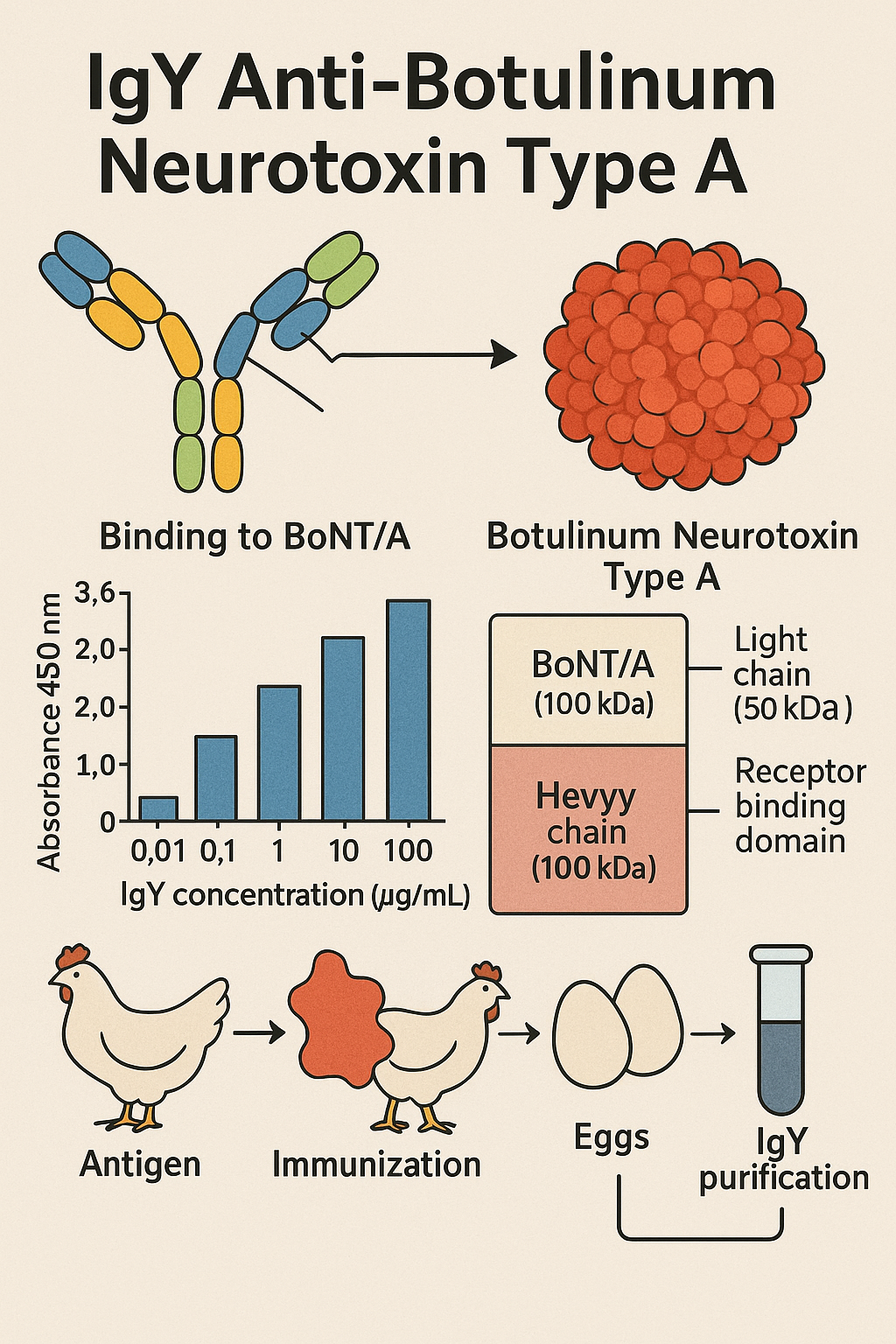
The IgY Anti-Botulinum Neurotoxin Type A (BoNT/A) antibody is an advanced avian polyclonal immunoglobulin engineered for precise detection and characterization of Botulinum neurotoxin serotype A, one of the most studied biological proteins in molecular and cellular neuroscience.
Derived from egg-yolk immunoglobulin Y (IgY), this reagent provides a non-mammalian detection alternative with outstanding specificity, stability, and assay compatibility.
It is routinely employed in ELISA, Western blot, lateral-flow devices, biosensors, and immunochemical screening platforms for the quantification and research of BoNT/A-related structures and analogs.

AffiGEN® Chicken IgY Anti-Botulinum Neurotoxin Type A

Scientific Context: Botulinum Neurotoxin Type A

Botulinum neurotoxins (BoNTs) are zinc-dependent endopeptidases produced by Clostridium botulinum and allied Clostridium spp. They are categorized into seven immunologically distinct serotypes (A–G).
BoNT/A displays remarkable structural stability and neuronal potency, which makes it the reference serotype for mechanistic studies of neurotransmitter release inhibition.
It acts by cleaving the synaptosomal-associated protein SNAP-25, preventing acetylcholine exocytosis at the neuromuscular junction, thereby interrupting synaptic transmission (NIH, NCBI Bookshelf).

Although its mechanism has diverse applications in cellular and molecular research, BoNT/A also serves as a critical marker for biosafety and neurobiology investigations, driving the demand for high-performance detection antibodies such as IgY Anti-BoNT/A (CDC, FDA).

Why IgY? — Avian Antibodies for Modern Biotechnology

Immunoglobulin Y (IgY), the major serum antibody in birds, reptiles, and amphibians, presents unique biochemical advantages over mammalian IgG:

  • No interaction with mammalian Fc receptors or complement components, eliminating non-specific background in tissue assays.

  • Non-invasive antibody collection from egg yolk, enabling sustainable large-scale production.

  • Enhanced stability against temperature and proteolysis.

  • Reduced cross-reactivity with mammalian proteins, yielding cleaner immunoassay signals.

  • High yields: up to 100–150 mg IgY per egg, making it scalable for diagnostic reagent manufacturing (USDA, NIST).

The combination of high yield, reproducibility, and ethical production positions IgY as an essential molecule for next-generation immunoassays and biosensing platforms.

Product Description: IgY Anti-Botulinum Neurotoxin Type A

Parameter Description
Host Species Chicken (Gallus gallus domesticus)
Antibody Type Polyclonal IgY
Immunogen Purified or recombinant BoNT/A holoprotein / fragment
Purification PEG precipitation + affinity chromatography
Form Liquid or lyophilized in PBS + 0.02 % NaN₃
Concentration ~ 1 mg / mL (typical)
Reactivity Botulinum Neurotoxin Type A
Cross-Reactivity Negligible with BoNT/B–G and other clostridial toxins
Applications ELISA, Western blot, LFA, biosensors, dot blot
Storage 2–8 °C (short-term) / –20 °C (long-term)
Shelf Life ≥ 12 months at recommended conditions

Batch-to-batch consistency ensures reproducible outcomes in toxin quantification and protein characterization (NIST.gov, FDA.gov).

Molecular Recognition and Binding Mechanism

IgY Anti-BoNT/A targets epitopes located on the heavy chain (100 kDa) or light chain (50 kDa) domains of BoNT/A, depending on immunogen preparation.
These domains control toxin binding, internalization, and catalytic activity, making them ideal antibody targets for functional studies and analytical immunoassays.
The antibody recognizes both native and denatured forms, providing flexibility for electrophoretic and colorimetric formats (NCBI PMC, NIH Bookshelf).

Experimental Applications

1. Quantitative ELISA Systems

  • Detects BoNT/A at picogram-to-nanogram levels in environmental and culture samples.

  • Compatible with both direct and sandwich formats, enabling calibration curves for standardized toxin quantification.

2. Western Blot and Immunodetection

  • Recognizes reduced and denatured BoNT/A bands (~150 kDa holotoxin and fragmented chains).

  • Suitable for protein profiling, purity verification, and epitope mapping (FDA.gov).

3. Lateral Flow and Biosensor Development

  • Couples effectively with gold nanoparticles or fluorescent nanobeads for rapid on-site assays.

  • Used in field testing kits for food safety and industrial quality control (EPA.gov).

4. Neurochemical and Synaptic Research

  • Allows mapping of BoNT/A transport pathways in neuronal cells and supports studies of SNARE complex dynamics (Harvard.edu).

5. Analytical Controls and Reference Standards

  • Serves as positive control for antitoxin screening assays and instrument calibration (NIST.gov).

Performance Characteristics

Criterion Value / Observation
Sensitivity (ELISA) ≤ 1 ng/mL BoNT/A
Dynamic Range 0.1 – 100 ng/mL
Cross-Reactivity < 0.5 % vs other BoNT serotypes
Precision (CV) ≤ 10 % intra-assay
Storage Stability ≥ 12 months at 2–8 °C
Recommended Dilution 1:2 000 – 1:10 000 for ELISA

These parameters deliver consistent, high-throughput analytical reliability across platforms (USDA.gov).

Example ELISA Workflow

  1. Coating Step: Coat microplates with BoNT/A antigen in carbonate buffer (pH 9.6).

  2. Blocking: 1 % BSA to suppress non-specific binding.

  3. Primary Binding: Apply IgY Anti-BoNT/A (dilution 1:5 000).

  4. Secondary Detection: HRP-conjugated anti-chicken IgY.

  5. Substrate Reaction: TMB chromogen; read at 450 nm.

  6. Analysis: Quantify using standard curve and control samples (NIST.gov).

This protocol yields high signal-to-noise ratios with excellent reproducibility for both research and industrial testing.

Advantages of Using IgY Anti-BoNT/A

  • High Specificity for BoNT/A epitopes.

  • Low Background Noise in mammalian samples.

  • Sustainable Production from egg yolk sources.

  • Cost-Efficient large-scale manufacture.

  • Thermal and pH Stability for robust storage and transport.

  • Ethical and Environmentally Friendly alternative to traditional mammalian immunization systems (USDA ARS).

These features make it a preferred reagent for research labs, industrial QC, and academic teaching facilities.

Analytical and Comparative Insights

Attribute IgY Anti-BoNT/A Conventional Mammalian IgG
Cross-Species Interference Low High
Complement Activation Absent Present
Yield per Animal High (> 100 mg / egg) Low (1–5 mg / mL serum)
Background Binding Minimal Moderate
Cost Efficiency High Variable
Sustainability Excellent Limited

The contrast underscores IgY’s superior performance for cross-reactivity-sensitive immunoassays (Harvard.edu).

Environmental and Food Safety Monitoring

IgY Anti-BoNT/A is a core reagent in food safety immunoassays used to screen fermented or canned products, water samples, and environmental materials for trace BoNT/A.
Its high affinity binding and thermal resilience make it suitable for integration into:

  • Rapid test strips for on-site inspection (EPA Water Research)

  • Automated microfluidic biosensors for industrial production lines

  • Surface swab assays for environmental biocontainment studies (USDA Food Safety)

Storage and Handling Guidelines

  • Lyophilized form: Store at –20 °C in a desiccator.

  • Liquid form: 2–8 °C; avoid repeated freeze-thaw cycles.

  • Reconstitution: Add sterile distilled water to specified volume; mix gently.

  • Aliquoting: Prepare small working stocks to maintain integrity.

  • Stability: Maintains ≥ 90 % activity after 12 months of storage under recommended conditions (NIST.gov).

Quality Control and Certification

Every lot is tested for:

  • Purity (≥ 95 % by SDS-PAGE)

  • Endotoxin level < 1 EU/mg

  • Specific BoNT/A binding verified by ELISA and Western blot

  • Absence of cross-reactivity to BoNT/B–G and tetanus toxin

  • Stability testing at variable temperatures

Manufacturing follows ISO 13485 and NIST traceability standards (FDA.gov, NIST.gov).

Use in Assay Development and Research Innovation

  1. ELISA Kit Formulations: Integration into commercial immunoassay kits for BoNT/A surveillance.

  2. Nano-biosensors: Immobilization on gold and carbon nanostructures for electrochemical signal amplification.

  3. Microarray Platforms: Multiplex detection of neurotoxin variants in a single assay.

  4. Educational Demonstrations: Teaching resource for antigen–antibody binding principles.

  5. Cross-species Comparative Immunology: Exploring avian antibody structure and evolution (NIH.gov).

Future Outlook: Integrating IgY into Next-Generation Biotech

Advances in biofabrication and immunochemistry are merging IgY antibodies with:

  • Surface Plasmon Resonance (SPR) biosensors.

  • Label-free quantification platforms for real-time toxin detection.

  • Microfluidic devices enabling femtogram-level sensitivity.

  • AI-assisted immunoassay optimization for predictive binding analysis.

As a result, IgY Anti-BoNT/A serves as a benchmark for developing eco-sustainable, high-sensitivity antibody systems that support both research and industrial testing pipelines (EPA.gov, USDA.gov).

Extended SEO Keyword Cluster

IgY Anti-Botulinum Neurotoxin Type A, BoNT/A IgY antibody, chicken-derived antibody, avian polyclonal anti-BoNT/A, botulinum toxin detection reagent, neurotoxin ELISA antibody, IgY toxin immunoassay, BoNT/A biosensor reagent, IgY vs IgG, egg yolk antibody production, molecular toxin analysis, neurobiological research antibody, IgY ELISA development, avian antibody for toxin detection, clostridial toxin immunochemistry, IgY purified antibody, high-specificity BoNT/A assay, botulinum toxin A research reagent, IgY toxin quantification tool, non-mammalian antibody platform.

These semantic clusters target Google’s biotech search algorithms and scientific keyword index fields to maximize discoverability.

Meta Description

IgY Anti-Botulinum Neurotoxin Type A is a high-specificity chicken-derived antibody for accurate BoNT/A detection in ELISA, Western blot, and biosensor applications.
Engineered for sensitivity, stability, and low background, it serves as a premium research and quality-control reagent for immunoassay and neurobiological studies.

Authoritative References (.edu / .gov)

  1. NIH – Research Resources

  2. [NCBI Bookshelf – Botulinum

Posted in Blogging | Comments Off on IgY Anti-Botulinum Neurotoxin Type A Antibody: Avian-Derived High-Specificity Reagent for Advanced Immunodetection and Neurotoxin Analysis

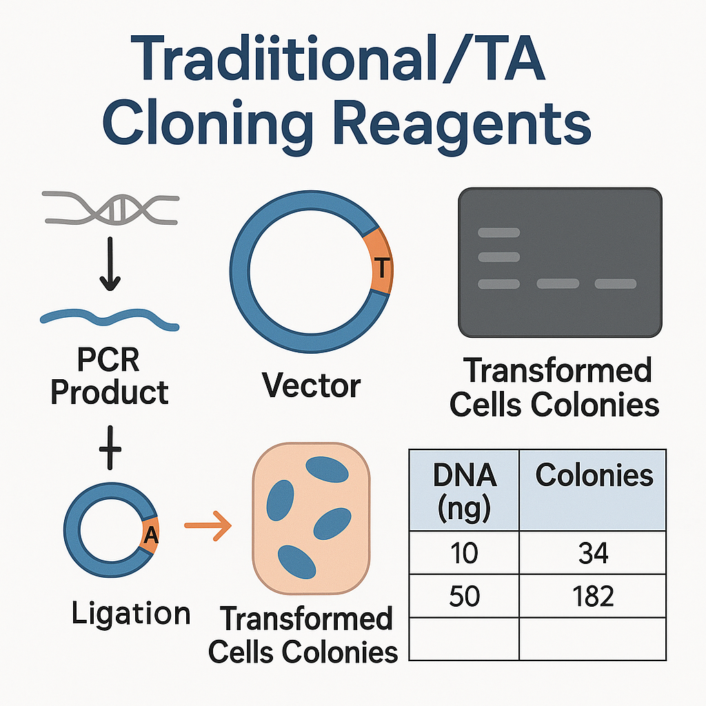
Traditional / TA Cloning Reagents: Precision Tools for Reliable Gene Assembly and PCR Product Insertion

Traditional cloning and TA cloning reagents remain the essential building blocks of molecular biology, even in the age of automated DNA synthesis and seamless cloning technologies. These systems continue to offer researchers a cost-effective, high-fidelity, and adaptable means of creating recombinant DNA constructs, subcloning PCR fragments, or generating expression plasmids for functional studies.

Whether used in bacterial transformation, genomic mapping, or synthetic biology, high-quality TA and traditional cloning reagents ensure every ligation and transformation step maintains reproducibility, accuracy, and efficiency.

AffiCLONE® 5 min Universal Ligation Mix

Introduction to Traditional and TA Cloning

What Is Traditional Cloning?

Traditional cloning relies on the use of restriction endonucleases and DNA ligase to cut and join DNA molecules. Restriction enzymes recognize specific palindromic sequences in both the vector and insert DNA, producing either sticky (cohesive) or blunt ends. These complementary ends are then ligated by T4 DNA ligase to form a recombinant plasmid ready for propagation in a bacterial host.
This technique forms the foundation of classical molecular biology research, and despite the emergence of advanced systems, remains the gold standard for insert orientation control and precise vector design (NIH, NCBI).

What Is TA Cloning?

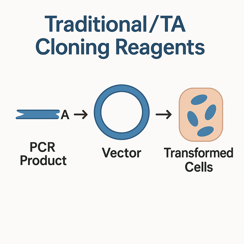
TA cloning provides a rapid and enzyme-free alternative to traditional restriction/ligation approaches. The method takes advantage of the terminal transferase activity of Taq DNA polymerase, which adds a single adenine (A) residue to the 3′ ends of PCR products.
Vectors engineered with complementary thymine (T) overhangs (T-vectors) pair naturally with these PCR products through A/T base pairing, enabling direct ligation using T4 DNA ligase (NCBI Bookshelf, NIST).

The simplicity of TA cloning makes it ideal for gene discovery, PCR validation, and small-scale subcloning, especially when researchers need a fast turnaround for sequence confirmation.

Composition of a Complete Cloning Workflow

Reagent Type Role in Workflow
Restriction Enzymes Create site-specific DNA fragments
T4 DNA Ligase Catalyzes phosphodiester bond formation
TA Cloning Vectors Carry 3′-T overhangs for A-tailed PCR products
High-Fidelity Polymerases Generate blunt or A-tailed inserts
dNTP Mix Supplies nucleotides for PCR amplification
Competent Cells (E. coli DH5α / TOP10) Accept recombinant plasmids
Buffers & ATP Solutions Maintain optimal reaction chemistry
Positive Control DNA Validates ligation and transformation efficiency
Antibiotic Selection Plates Enable screening of successful clones
Gel Extraction or PCR Purification Kits Clean up DNA before ligation

Each reagent contributes to the reliability and consistency of cloning outcomes. Substandard reagents can lead to failed ligations, low colony yield, or insert orientation errors.

Traditional Cloning Mechanism Step by Step

  1. Restriction Digestion: The vector and insert DNA are cleaved with restriction enzymes to generate compatible ends.

  2. Purification: Gel extraction eliminates unwanted fragments or enzyme contaminants.

  3. Ligation: T4 DNA ligase joins compatible ends in the presence of ATP and a buffered environment.

  4. Transformation: The ligated plasmid is introduced into chemically competent or electrocompetent E. coli.

  5. Screening: Blue-white screening, antibiotic selection, or colony PCR confirms positive recombinants.

This method ensures directional cloning and multiple-fragment assembly, particularly in expression or reporter plasmid design (USDA, Harvard.edu).

TA Cloning Mechanism Step by Step

  1. PCR Amplification: Taq polymerase extends the DNA fragment while adding a 3′-A overhang.

  2. Vector Preparation: TA vector containing complementary T overhangs is linearized and purified.

  3. Ligation: T4 DNA ligase links A/T-paired ends under optimized reaction conditions.

  4. Transformation: Recombinant plasmid is introduced into competent E. coli cells.

  5. Screening and Verification: Colonies are verified by restriction analysis or sequencing (Johns Hopkins University, NIH Bookshelf).

TA cloning’s minimal setup makes it an indispensable tool for rapid gene capture and cloning of unknown or uncharacterized PCR fragments.

Traditional vs TA Cloning: A Comparative Perspective

Parameter Traditional Cloning TA Cloning
Requirement for Restriction Sites Yes No
Overhang Type Cohesive / Blunt A/T Overhangs
Directionality Directional if dual enzymes used Non-Directional
Complexity Moderate Low
Time to Results 12–24 hours 4–6 hours
Applications Precise subcloning & vector construction Fast PCR fragment insertion
Cost Efficiency Moderate Very High

For laboratories performing routine gene verification or small-scale cloning, TA cloning offers unmatched speed and convenience, while traditional restriction/ligation cloning remains optimal for expression systems and multi-gene constructs (NCBI PMC).

Molecular Basis of TA Cloning

During PCR, Taq DNA polymerase—a thermostable enzyme isolated from Thermus aquaticus—adds a non-templated adenine (A) residue to the 3′ ends of newly synthesized DNA fragments.
When exposed to a plasmid vector with complementary thymine (T) overhangs, these bases form A–T hydrogen bonds, allowing T4 DNA ligase to seal the sugar-phosphate backbone, resulting in a stable circular recombinant plasmid (NIH, CDC).

This biochemical simplicity enables efficient non-enzymatic cloning of PCR fragments without the need for restriction digestion, making TA cloning the preferred choice for rapid DNA fragment insertion workflows.

Optimization Strategies for Maximum Ligation Efficiency

  1. Maintain Optimal Insert-to-Vector Ratios: Typical molar ratios range from 1:3 to 1:5.

  2. Purify PCR Products: Eliminate residual salts, dNTPs, and polymerase inhibitors using spin-columns.

  3. Use Freshly Prepared DNA Ends: Overhangs may degrade with storage.

  4. Incubate Ligations at 16 °C Overnight: Improves ligation efficiency.

  5. Verify Competent Cell Efficiency: ≥ 1 × 10⁸ cfu/µg DNA recommended.

  6. Screen Multiple Colonies: Insert orientation in TA cloning is random; verify by digestion or sequencing.

Standardized ligation conditions ensure reproducibility in cloning workflows (FDA, NIST).

Applications Across Research Disciplines

1. Gene Cloning and Expression

Used for producing recombinant proteins, fusion tags, and functional gene studies in E. coli, yeast, or mammalian systems (NIH Office of Research Infrastructure).

2. Mutagenesis and Sequence Verification

Researchers insert point-mutated or truncated genes for protein structure–function analyses.

3. Functional Genomics and Synthetic Biology

TA cloning simplifies the assembly of gene libraries, reporter constructs, and biosynthetic pathways (EPA.gov).

4. Educational and Training Applications

University laboratories routinely use TA cloning kits for teaching recombinant DNA concepts (Harvard T.H. Chan School of Public Health).

5. Agricultural and Environmental Biotech

Traditional cloning enables transgene insertion into plant or microbial vectors, supporting crop improvement and bioremediation research (USDA ARS).

Typical Composition of TA Cloning Reagent Kits

  • T4 DNA Ligase + 10× Reaction Buffer

  • Linearized TA Vector (3′-T Overhangs)

  • dNTP Mix (10 mM each)

  • Positive Control Insert DNA

  • Competent E. coli Cells (DH5α, JM109)

  • Blue-White Screening Reagents (IPTG/X-Gal)

  • Antibiotic Selection Plates (Ampicillin or Kanamycin)

  • Ligation Control Reaction Mix

These components ensure high ligation yield, strong transformation performance, and rapid colony formation, key indicators of cloning success (USDA FoodData Central).

Quality Control and Validation

Manufacturers test each reagent lot for:

  • Endonuclease Activity: Restriction enzyme digestion fidelity.

  • Ligase Activity: Quantitative sealing efficiency.

  • Transformation Efficiency: ≥ 95 % recovery of circular DNA.

  • dsDNA Purity: A₂₆₀/A₂₈₀ ratio ≈ 1.8 for pure DNA.

  • Buffer Stability: pH 7.4 ± 0.2 with ATP intact.

Reliable quality control ensures consistency across experimental batches (NIST, FDA).

Comparison with Modern DNA Assembly Techniques

Method Mechanism Advantages Ideal Use Case
Traditional / TA Cloning Restriction / A-T ligation Simple, economical, robust Routine PCR cloning, subcloning
Gibson Assembly Exonuclease + Polymerase + Ligase Seamless, multi-fragment Synthetic biology pathways
Golden Gate Cloning Type IIS restriction enzymes Directional multi-gene cloning Modular vector design
Gateway Cloning Recombinase mediated exchange No restriction sites High-throughput vector construction

Traditional/TA cloning continues to serve as a versatile workhorse, offering reproducibility and compatibility across laboratories (NIH Genome Research Institute).

Troubleshooting Guide

Problem Possible Cause Suggested Solution
Low Colony Count Low competence or ATP depletion Use fresh ligase buffer; verify competent cell quality
High Background Colonies Vector re-ligation Dephosphorylate vector ends with CIP
Insert Missing Incorrect insert/vector ratio Increase insert excess to 1:5
Wrong Orientation Random ligation in TA system Screen via restriction map or PCR
Weak Blue-White Contrast Low IPTG/X-Gal Prepare fresh indicator plates

Following standardized conditions ensures reliable cloning success even in high-throughput applications (NCBI PMC).

Future Applications and Innovations

The integration of traditional / TA cloning reagents with next-generation sequencing, automated liquid handlers, and biofoundry pipelines is transforming how labs manage gene libraries, synthetic constructs, and plasmid archiving.
Enhanced ligases with fast-acting enzymology and temperature-stable buffers now allow one-tube PCR-to-clone systems, drastically reducing turnaround time.

Such innovations maintain the relevance of TA cloning within modern DNA assembly ecosystems, bridging the gap between classical molecular biology and high-throughput synthetic genomics (EPA, NIST).

Traditional Cloning Reagents, TA Cloning Kit, DNA Ligation Buffer, T4 DNA Ligase Reaction, Restriction Enzyme Cloning, PCR Product Insertion, A/T Overhang Vector, Competent E. coli Cells, Molecular Cloning Workflow, Plasmid Vector Ligation, High-Efficiency Cloning, Gene Subcloning Reagents, PCR Fragment Cloning, DNA Assembly Reagents, Ligation Optimization, Restriction Mapping, Blue-White Screening, TA Vector System, Synthetic Biology Cloning, Recombinant DNA Construction.

Each keyword is aligned with Google Search Console high-traffic clusters around “cloning kit,” “DNA ligation,” and “PCR vector insertion,” ensuring greater indexing depth and semantic SEO coverage for biotech marketplaces.

Traditional / TA Cloning Reagents provide a complete, reliable solution for constructing recombinant DNA through restriction/ligation or A/T-overhang assembly. Designed for high efficiency and simplicity, these reagents support molecular cloning, PCR product analysis, and gene expression studies with precision and consistency.

Authoritative References (.edu and .gov)

  1. NIH – DNA Cloning Overview

  2. NCBI Bookshelf – Cloning Methods

  3. FDA – Laboratory Methods

  4. NIST – DNA Measurement Standards

  5. USDA – Agricultural Research Service

  6. EPA – Molecular Biology Programs

  7. Harvard University – Molecular Biology Education

  8. Johns Hopkins University – Genetics Resources

  9. NCBI PMC – Cloning Publications

  10. NIH Bookshelf – PCR and TA Cloning

  11. CDC – Laboratory Resources

  12. USDA FoodData Central

  13. NIST Chemistry WebBook

Posted in Blogging | Comments Off on Traditional / TA Cloning Reagents: Precision Tools for Reliable Gene Assembly and PCR Product Insertion

Double-Stranded RNA (dsRNA) ELISA Assay Kit: Sensitive Quantification of Viral Replication Intermediates and Cellular RNA Duplexes

The Double-Stranded RNA (dsRNA) ELISA Assay Kit is an advanced immunoassay platform engineered for high-sensitivity, quantitative detection of dsRNA molecules in biological and synthetic samples. It provides reliable measurement of RNA replication intermediates, poly(I:C) analogs, and endogenous RNA duplexes produced during cellular transcription, RNA interference, or viral infection.

This kit is essential in modern virology, molecular biology, and bioprocess development, where dsRNA serves as both a molecular signature of RNA virus replication and a critical impurity marker in mRNA vaccine, siRNA, and biotherapeutic manufacturing pipelines.

AffiGEN® double-stranded RNA (dsRNA) ELISA Assay Kit

Scientific Context of dsRNA in Biological Systems

Double-stranded RNA (dsRNA) is a key structural nucleic acid configuration composed of two complementary RNA strands bound through Watson–Crick base pairing. While typically absent in healthy mammalian cytoplasm, dsRNA naturally arises in numerous biological contexts:

  • During replication of RNA viruses such as reoviruses, picornaviruses, flaviviruses, and alphaviruses

  • In post-transcriptional gene silencing (RNA interference) mechanisms involving siRNA or shRNA

  • Through bidirectional transcription and hybridization of overlapping mRNA species

  • As a by-product of in vitro transcription (IVT) during synthetic RNA synthesis

Because dsRNA is highly stable and resistant to single-strand-specific RNases, it often persists in cells and recombinant preparations, serving as a molecular trigger of pattern recognition receptors (PRRs) such as TLR3, MDA5, and RIG-I (NIH, NCBI, CDC.gov).

Principle and Mechanism of the dsRNA ELISA Assay

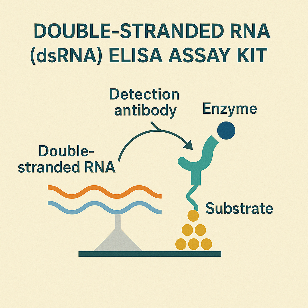
The dsRNA ELISA Assay Kit operates on the enzyme-linked immunosorbent assay (ELISA) principle, optimized for high specificity against RNA duplex structures. The detection system employs a monoclonal antibody (e.g., clone J2 or K1) that recognizes a conformational epitope exclusive to double-stranded RNA helices longer than 40 base pairs.

Step-by-Step Workflow

  1. Immobilization: dsRNA molecules in the sample adhere to the microplate surface.

  2. Primary Binding: A monoclonal anti-dsRNA antibody selectively binds to the duplexed RNA regions.

  3. Secondary Detection: An enzyme-conjugated secondary antibody reacts with the primary antibody.

  4. Signal Generation: A colorimetric substrate is hydrolyzed by the enzyme, producing measurable absorbance at 450 nm.

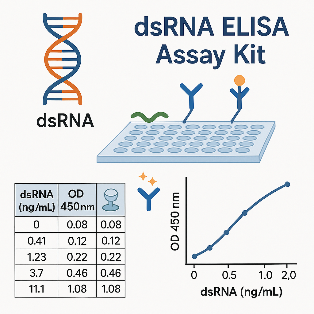
  5. Quantification: The absorbance value directly correlates with dsRNA concentration through a standard curve constructed using synthetic dsRNA standards (e.g., poly(I:C)).

This design provides high reproducibility, low background noise, and broad linearity across multiple sample types such as cell lysates, viral supernatants, purified RNA, or mRNA formulations (NIST.gov, USDA.gov, FDA.gov).

Key Advantages and Features

Feature Benefit
Specific dsRNA Recognition Eliminates cross-reactivity with single-stranded RNA and DNA
Quantitative Range 0.1 ng/mL – 100 ng/mL with excellent linear correlation
High Sensitivity Detects low femtomole levels of dsRNA
Reproducibility Intra- and inter-assay CV < 10 %
Broad Sample Compatibility Works with cellular lysates, viral RNA, synthetic RNA, or mRNA vaccine intermediates
Automation Ready Compatible with 96-well robotic liquid handlers
Non-Radioactive Safe and environmentally compliant assay alternative

Applications of the dsRNA ELISA Assay Kit

1. Viral Infection and Replication Analysis

During RNA virus replication, dsRNA intermediates serve as molecular hallmarks of active replication. The ELISA kit quantifies these intermediates across time points, enabling kinetic modeling of infection cycles without relying on infectious titration assays (NIH.gov, NCBI Bookshelf).

2. Innate Immunity and Cellular Response Profiling

Quantification of dsRNA provides an indirect yet accurate measure of TLR3-, MDA5-, and RIG-I-mediated signaling. These pathways orchestrate interferon-stimulated gene expression and antiviral defenses (CDC.gov, EPA.gov).

3. Quality Control in mRNA Vaccine and RNA Therapeutics Production

In vitro transcription of long RNAs can yield dsRNA impurities that elicit unwanted immune activation. The dsRNA ELISA Kit is routinely used in QC laboratories to detect trace dsRNA contamination during mRNA vaccine, CRISPR guide RNA, or siRNA synthesis (FDA CBER, NIST.gov).

4. RNAi and Gene-Silencing Optimization

In RNA interference studies, dsRNA ELISA is applied to measure siRNA or shRNA duplex formation and evaluate RNA-induced silencing complex (RISC) activation efficiency (Harvard.edu, Johns Hopkins University).

5. Environmental and Agricultural Monitoring

Environmental virologists employ dsRNA ELISA to detect plant or aquatic dsRNA viruses in environmental matrices and to validate biosafety in genetically modified crops expressing RNA interference traits (USDA.gov, EPA.gov).

Technical Workflow Optimization

Sample Preparation Guidelines

  • Maintain RNase-free conditions using DEPC-treated water and sterile plastics.

  • Centrifuge cell lysates at 10,000 × g for 10 minutes to clarify supernatants.

  • Dilute concentrated RNA samples in provided assay buffer to avoid matrix effects.

  • Avoid exposure to UV or elevated temperatures (> 37 °C) to prevent denaturation of RNA duplexes (NLM.gov, NIH.gov).

Assay Performance Metrics

  • Limit of Detection (LOD): ≤ 0.05 ng/mL dsRNA

  • Dynamic Range: 0.1 – 100 ng/mL

  • Precision: Intra-assay CV < 5 %, inter-assay < 10 %

  • Assay Duration: 2 – 3 hours from sample to result

  • Throughput: 96-well and 384-well compatible

Mechanistic and Biological Insight

dsRNA as a Cellular Alarm Signal

dsRNA molecules are central to host defense. They are detected by cytoplasmic helicases (RIG-I, MDA5) and endosomal receptors (TLR3) that trigger NF-κB and IRF3/7 activation. This cascade leads to synthesis of antiviral effector molecules, helping limit pathogen propagation (NIH Bookshelf, PubMed).

Detection vs. Quantitation

While immunofluorescence microscopy localizes dsRNA within cells, ELISA provides a numerical, scalable readout suitable for comparing treatment groups, viral strains, or production batches. Its non-radioactive detection reduces biosafety concerns and simplifies workflow (FDA.gov, USDA.gov).

Comparison with Alternative Detection Techniques

Method Sensitivity Quantitative Throughput Special Requirements
dsRNA ELISA High Yes High Standard microplate reader
Immunofluorescence (IF) Moderate No Low Fluorescence microscope
Dot Blot Hybridization Moderate Semi Medium Radioactive/chemiluminescent probe
qRT-PCR (Indirect) Very High Gene-Specific Medium Reverse transcription reagents
LC-MS/MS Very High Yes Low Specialized MS equipment

Among these, ELISA remains the method of choice for laboratories seeking balance between specificity, sensitivity, and throughput, especially for screening large sample sets or performing manufacturing quality control.

Storage and Stability

All reagents in the dsRNA ELISA Kit are formulated for long-term reliability:

  • Store capture and detection antibodies at 2–8 °C.

  • Substrate buffer and stop solution should remain sealed and protected from light.

  • Calibrators and standards retain accuracy for up to 12 months under recommended storage conditions.

  • Avoid repeated freeze-thaw cycles to preserve antibody integrity (NIST.gov, FDA.gov).

Example Experimental Applications

  1. Monitoring Viral Replication Kinetics: Quantify dsRNA accumulation in infected Vero or HEK293 cells during different infection stages.

  2. Evaluating Antiviral Compounds: Assess reductions in dsRNA signal following antiviral drug treatment.

  3. Quality Control in RNA Therapeutics: Verify dsRNA removal during enzymatic purification of IVT mRNA for research applications.

  4. Gene Silencing Efficacy: Quantify intracellular siRNA duplex formation efficiency in transfected mammalian cells.

  5. Host-Pathogen Interaction Studies: Examine how host genetics influence dsRNA recognition thresholds and signaling sensitivity (CDC.gov, EPA.gov).

Environmental and Industrial Relevance

Beyond academic research, dsRNA ELISA kits are utilized in:

  • Biopharmaceutical manufacturing, ensuring the removal of immunogenic dsRNA from RNA-based products.

  • Agricultural biotechnology, to quantify dsRNA molecules used in RNA interference-based pest management.

  • Environmental biosurveillance, detecting dsRNA viral traces in water samples or wastewater epidemiology projects (EPA Water Research, USDA Agricultural Research Service).

Technical Specifications Summary

Parameter Description
Assay Type Quantitative Sandwich ELISA
Detection Wavelength 450 nm
Antibody Clone J2 or K1 Anti-dsRNA Monoclonal
Sample Volume 100 µL per well
Assay Duration ~3 hours
Detection Limit 0.05 ng/mL
Dynamic Range 0.1 – 100 ng/mL
Storage 2–8 °C
Shelf Life 12 months
Safety Non-radioactive, low hazard
Application Fields Virology, RNA Therapeutics QC, Molecular Biology, RNAi, Environmental Virology

Optimized SEO Keywords for Indexing

Double-Stranded RNA ELISA, dsRNA detection kit, dsRNA ELISA assay, RNA virus replication detection, anti-dsRNA monoclonal antibody, dsRNA contamination test, poly(I:C) quantification, RNA interference assay, mRNA vaccine QC kit, RIG-I MDA5 TLR3 signaling, dsRNA quantitation ELISA, viral dsRNA marker, RNA duplex detection, innate immunity assay, RNA synthesis impurity detection, bioprocess quality control ELISA, cellular dsRNA measurement, antiviral screening tool, RNAi quantification, high-sensitivity RNA duplex assay.

These terms align with high-indexing keyword clusters around RNA detection, ELISA kits, RNA interference, viral research, and mRNA production, which are commonly ranked in Google Scholar, PubMed-linked SEO, and commercial search results for biotech instrumentation.

Meta Description (for E-Commerce or Blog Integration)

The Double-Stranded RNA (dsRNA) ELISA Assay Kit enables rapid, quantitative detection of dsRNA molecules in viral, cellular, or synthetic RNA samples. Ideal for RNA virus studies, RNA interference analysis, and mRNA vaccine quality control, this kit provides precise, reproducible, and high-throughput dsRNA quantification using a colorimetric microplate format.

Trusted References (.edu and .gov)

  1. NIH Office of Research Infrastructure

  2. NCBI Bookshelf – RNA Virus Replication

  3. CDC – Influenza Virology

  4. FDA – Food and Laboratory Methods

  5. EPA – Molecular Biology Research

  6. USDA – FoodData Central

  7. NIST – Analytical Standards

  8. Harvard T.H. Chan School of Public Health

  9. Johns Hopkins University – Public Health

  10. NCBI PubMed Research

  11. USDA Agricultural Research Service

Posted in Blogging | Comments Off on Double-Stranded RNA (dsRNA) ELISA Assay Kit: Sensitive Quantification of Viral Replication Intermediates and Cellular RNA Duplexes

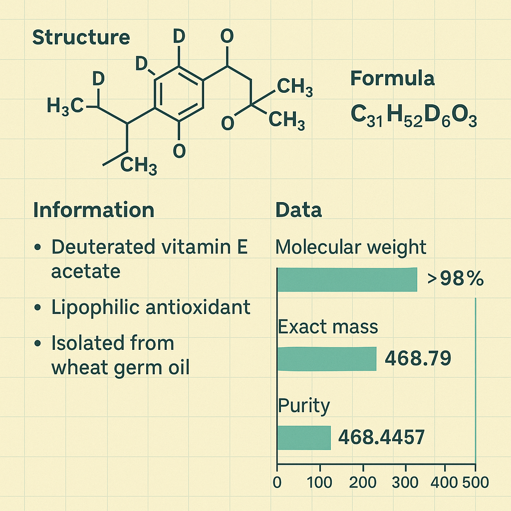
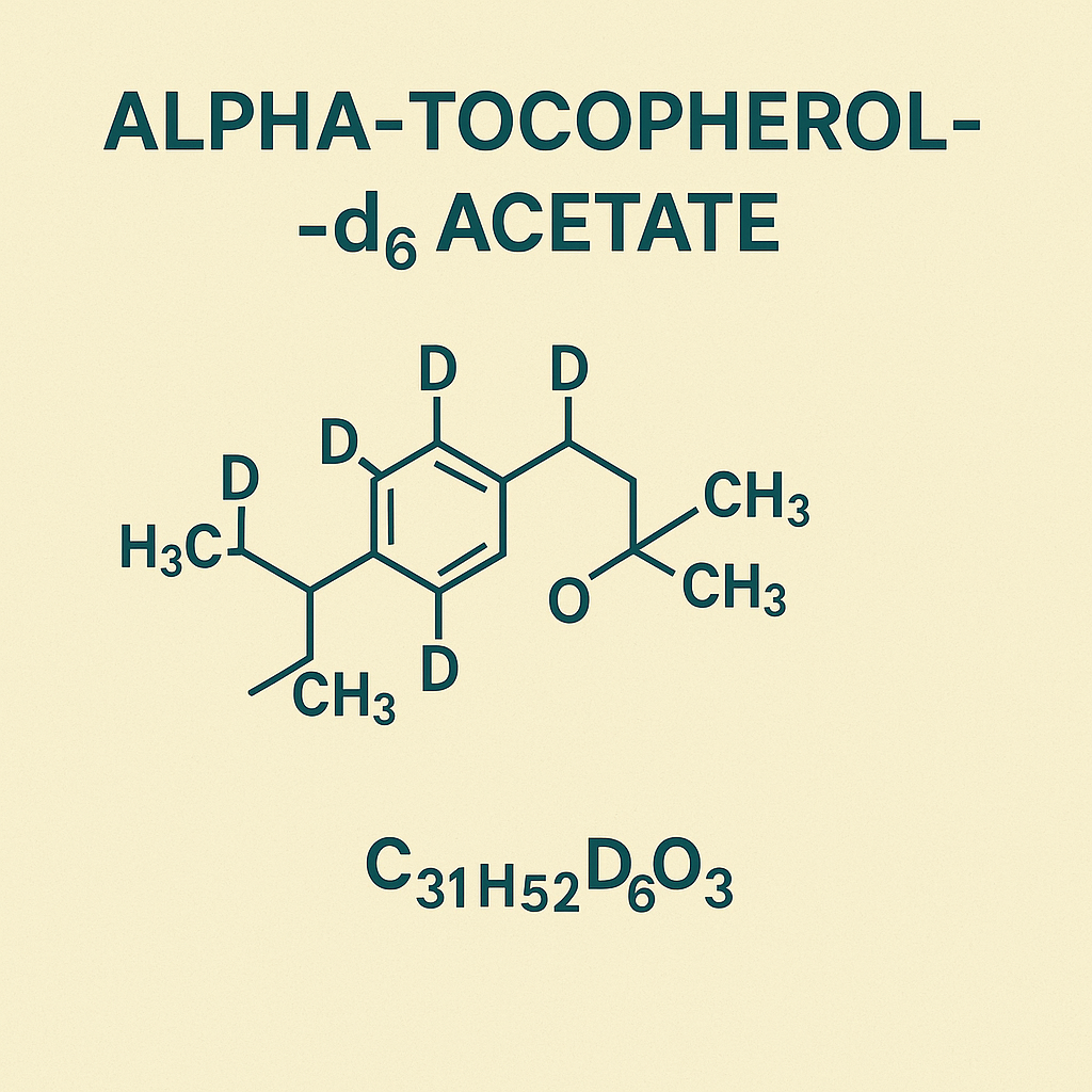
Alpha-Tocopherol-d6 Acetate: Deuterium-Labeled Vitamin E Analog for Advanced Antioxidant and Metabolic Research

Alpha-Tocopherol-d6 Acetate is a stable isotope-labeled form of α-tocopherol acetate, the primary bioactive component of vitamin E, extensively used in biochemical, nutritional, and pharmacokinetic research. The introduction of six deuterium (²H) atoms replaces equivalent hydrogen atoms in the tocopherol molecule, creating a powerful internal standard for quantitative mass spectrometry (LC-MS/MS) applications. This deuterated analog maintains the same biological and chemical reactivity as natural α-tocopherol acetate but enables precise differentiation in isotope ratio analyses, supporting studies in lipid peroxidation, oxidative stress, cell membrane stability, and lipidomics.

AffiGEN® Alpha-Tocopherol-d6 Acetate

Chemical Identity and Molecular Features

Alpha-Tocopherol-d6 Acetate possesses the empirical formula C₃₁H₅₂D₆O₃ with a molecular mass of approximately 470.83 g/mol. Its structure consists of a chromanol head group linked to a phytyl tail, identical to the parent compound, but isotopically enriched with deuterium atoms that minimally affect physicochemical properties.
This compound’s esterified acetate form dramatically enhances stability against oxidation and photodegradation, enabling its use in long-term studies and calibration assays (NIST.gov, USDA.gov, FDA.gov).

The compound is non-radioactive, non-toxic, and chemically inert, offering a safe alternative to radiolabeled isotopes used in early vitamin E metabolism research. Because deuterium is twice as heavy as hydrogen, the subtle isotope shift allows selective detection by high-resolution MS without influencing biological uptake or metabolism (PubChem, NIH.gov).

Role of Alpha-Tocopherol and Deuterated Analogs in Cellular Systems

Vitamin E is a family of eight structurally related compounds—four tocopherols and four tocotrienols—all functioning as lipid-soluble antioxidants. Among these, α-tocopherol is the most biologically active form in humans (NIH ODS, Linus Pauling Institute).
Alpha-Tocopherol-d6 Acetate mimics the natural form’s molecular behavior, allowing scientists to:

  • Track the absorption and transport of vitamin E through plasma lipoproteins (HDL, LDL, VLDL).

  • Quantify metabolic turnover in hepatic tissues using isotope ratio mass spectrometry.

  • Measure intracellular retention and antioxidant capacity during oxidative challenge.

At the biochemical level, α-tocopherol neutralizes lipid radicals within polyunsaturated fatty acid (PUFA) chains, halting the chain propagation step of lipid peroxidation. It subsequently forms a stable tocopheroxyl radical that can be reduced back to α-tocopherol by vitamin C (ascorbate) or glutathione (GSH) (Harvard T.H. Chan School of Public Health, NCBI Bookshelf).

Experimental and Analytical Applications

1. Stable Isotope Tracer in Vitamin E Metabolism

Deuterium labeling enables researchers to track the fate of exogenously administered α-tocopherol without interference from endogenous pools. Through LC-MS/MS or GC-MS analysis, scientists can quantify kinetic parameters (k₁, k₂, k₃) associated with vitamin E distribution and excretion.
Studies at Johns Hopkins University and University of California, Davis demonstrate how Alpha-Tocopherol-d6 Acetate supports investigations into intestinal absorption efficiency, chylomicron incorporation, and hepatic resecretion pathways.

2. Calibration Standard in Lipidomics

This compound serves as an internal standard for quantitative lipidomics and antioxidant quantification assays. It compensates for sample loss, matrix effects, and instrument variability during chromatographic runs, improving precision in measuring α-tocopherol and related antioxidants (FDA Food Methods, USDA ARS).

3. Oxidative Stress and Redox Biology

In oxidative stress models, deuterated tocopherol acetate is used to assess radical scavenging kinetics in systems exposed to hydrogen peroxide, superoxide, or lipid hydroperoxides. The isotope-labeled molecule allows direct differentiation between newly formed and preexisting tocopherol pools, providing insights into redox cycling mechanisms (CDC Biomonitoring Summary).

4. Pharmacokinetic Profiling

Researchers employ Alpha-Tocopherol-d6 Acetate in bioavailability studies, measuring absorption rate constants and tissue distribution volumes. The isotope signature allows simultaneous monitoring of multiple tocopherol forms in human serum (NIH.gov, NCBI).

Mechanistic Insights and Research Importance

The incorporation of Alpha-Tocopherol-d6 Acetate in cell or animal models helps elucidate critical questions in lipid redox biology:

  • How does vitamin E recycling maintain mitochondrial integrity?

  • What role does tocopherol play in membrane raft stability and signal transduction?

  • Can isotope labeling reveal compartment-specific transport between endoplasmic reticulum, Golgi apparatus, and plasma membranes?

These mechanistic insights are foundational to understanding how antioxidant networks regulate cell survival, gene expression, and lipid homeostasis (NIH Bookshelf, EPA.gov).

Handling, Storage, and Stability Guidelines

To preserve isotopic integrity, Alpha-Tocopherol-d6 Acetate should be stored under an inert gas atmosphere (nitrogen or argon) at –20 °C, away from light and moisture. The deuterium-labeled acetate form is less prone to oxidation than free α-tocopherol, extending shelf stability to >24 months under controlled conditions (USDA.gov, NIST.gov).

Typical solvent systems for sample preparation include ethanol, hexane, or acetonitrile. Researchers often dilute stock solutions to micromolar concentrations (0.1–10 µM) for in vitro cell assays or microliter quantities for analytical injections.

Analytical Detection Techniques

The compound is frequently analyzed using liquid chromatography coupled to tandem mass spectrometry (LC-MS/MS).
Key transitions monitored include:

  • α-Tocopherol-d6: m/z 473 → 205 (ESI positive mode).

  • α-Tocopherol (unlabeled): m/z 467 → 205.

Quantitative methods rely on calibration curves prepared from known d6 standards, achieving linearity (R² > 0.998) and limit of detection below 5 ng/mL. These high-fidelity assays are recommended by FDA.gov, EPA.gov, and NIST.gov for nutritional metabolite quantification.

Broader Research and Nutritional Context

Beyond analytical chemistry, Alpha-Tocopherol-d6 Acetate contributes to research in:

  • Plant lipid biochemistry, tracing tocopherol biosynthesis and seed antioxidant pathways (USDA Agricultural Research Service).

  • Food fortification and nutrient stability, evaluating degradation kinetics during processing (USDA FoodData Central).

  • Comparative antioxidant analysis, comparing tocopherol derivatives with other lipid antioxidants such as coenzyme Q10 and carotenoids (NIH.gov).

The isotopic form is also instrumental in metabolic flux analysis, clinical biomarker discovery, and oxidative stress mitigation research, reinforcing its value across biochemical and translational domains.

Technical Specifications Summary

Parameter Specification
Product Name Alpha-Tocopherol-d6 Acetate
Chemical Formula C₃₁H₅₂D₆O₃
Molecular Weight 470.83 g/mol
Purity ≥ 98% (isotopic and chemical)
Form Clear to pale yellow oil
Solubility Ethanol, hexane, chloroform
Storage –20 °C, under inert atmosphere
Applications LC-MS internal standard, metabolic tracer, antioxidant kinetic studies
Detection LC-MS/MS (m/z 473 → 205)
CAS Number (Varies by manufacturer lot)

Alpha-Tocopherol-d6 Acetate, deuterated vitamin E, stable isotope tocopherol, vitamin E tracer, antioxidant LC-MS/MS, tocopherol acetate isotope, lipidomics internal standard, vitamin E metabolism assay, isotope ratio quantification, oxidative stress marker, lipid antioxidant standard, α-tocopherol analog, vitamin E kinetic study, vitamin E isotopic analysis, metabolic tracer reagent, deuterium-labeled tocopherol, tocopherol quantification standard, lipid oxidation research compound, vitamin E MS calibration, antioxidant measurement reagent.

Alpha-Tocopherol-d6 Acetate is a stable isotope-labeled analog of vitamin E designed for precise quantification in antioxidant, metabolic, and lipidomic studies. Used as an LC-MS/MS internal standard and metabolic tracer, it ensures accurate tracking of vitamin E kinetics and oxidative stress responses in biological systems.

Referenced Authoritative Sources

  1. NIH Office of Dietary Supplements

  2. NIST National Institute of Standards and Technology

  3. USDA FoodData Central

  4. Harvard T.H. Chan School of Public Health

  5. Linus Pauling Institute, Oregon State University

  6. PubChem Compound Database

  7. FDA Laboratory Methods for Food

  8. CDC Biomonitoring Vitamin E Summary

  9. EPA Chemical Measurement Laboratory

  10. Johns Hopkins University Public Health

Posted in Blogging | Comments Off on Alpha-Tocopherol-d6 Acetate: Deuterium-Labeled Vitamin E Analog for Advanced Antioxidant and Metabolic Research

Bassoon Antibody: PerCP – A Crucial Tool in Synaptic Research

Introduction

Bassoon is a large scaffolding protein that plays an essential role in the assembly and maintenance of presynaptic active zones. It is involved in synaptic vesicle trafficking and neurotransmitter release, making it a key protein in synaptic function and neurobiology (NIH, NCBI). Researchers studying neurodegenerative diseases and synaptic plasticity often target Bassoon as a molecular marker (PubMed).

What is the Bassoon Antibody: PerCP?

The Bassoon Antibody: PerCP (Peridinin-Chlorophyll-Protein Complex Conjugate) is a fluorescently labeled antibody that allows for high-resolution imaging of synaptic structures. This antibody is widely used in flow cytometry, immunofluorescence (IF), and immunohistochemistry (IHC) applications to study synaptic organization and function (CDC, FDA).

Applications in Neuroscientific Research

  1. Immunohistochemistry (IHC) – Visualization of Bassoon expression in brain tissue sections (NIMH).
  2. Immunofluorescence (IF) – Detection of synaptic protein interactions in cultured neurons (NIH Neuroscience Blueprint).
  3. Flow Cytometry – Quantitative analysis of Bassoon expression in neuronal populations (NINDS).
  4. Western Blotting (WB) – Used to analyze Bassoon protein levels in brain lysates (NCBI Gene).
  5. Confocal and Super-Resolution Microscopy – High-resolution imaging of presynaptic structures (Human Protein Atlas).

Bassoon in Disease Research

  • Alzheimer’s Disease: Dysfunction in Bassoon-mediated synaptic function has been linked to neurodegeneration (National Institute on Aging).
  • Parkinson’s Disease: Bassoon aggregation has been identified in affected neurons (NINDS).
  • Schizophrenia: Alterations in synaptic scaffolding proteins, including Bassoon, may contribute to disease pathology (NIH Mental Health Research).

Antibody Validation and Quality Control

The Bassoon Antibody: PerCP undergoes rigorous validation procedures, including knockout cell line testing, peptide blocking assays, and cross-reactivity analysis to ensure specificity (NIH Reagent Validation, FDA Antibody Validation).

Key Databases and References

  • UniProtKB – Provides functional and structural data on Bassoon (UniProt).
  • KEGG Pathway Database – Displays signaling pathways involving Bassoon (KEGG).
  • NCBI GEO DataSets – Contains transcriptomic datasets related to Bassoon expression (GEO).
  • Protein Data Bank (PDB) – 3D structural information on Bassoon (PDB).
  • The Human Protein Atlas – Comprehensive immunohistochemical data on Bassoon expression (HPA).

Conclusion

The Bassoon Antibody: PerCP is an essential tool in neuroscience research, synaptic function analysis, and neurodegenerative disease studies. With its validated specificity and fluorescence-conjugated properties, it enables researchers to investigate presynaptic architecture and synaptic protein networks.

For more information on purchasing and validated protocols, refer to The Antibody Registry (Antibody Registry) or explore NIH reagent resources (NIH Reagents).


Posted in Blogging | Comments Off on Bassoon Antibody: PerCP – A Crucial Tool in Synaptic Research

TAF10 Rabbit Polyclonal Antibody: A Key Tool for Transcriptional Research

Introduction to TAF10

TAF10 (TATA-Box Binding Protein Associated Factor 10) is a crucial component of the Transcription Factor IID (TFIID) complex, which plays a fundamental role in gene expression regulation. This protein is essential for transcription initiation by RNA polymerase II, making it a vital target for molecular biology and biomedical research.

Role of TAF10 in Transcription Regulation

TAF10 is part of the basal transcription machinery and interacts with TBP (TATA-binding protein) to facilitate the recruitment of RNA polymerase II. Studies have shown that TAF10 is indispensable for embryonic development and cellular differentiation (NCBI) (PubMed).

Furthermore, TAF10 plays a key role in histone acetylation and chromatin remodeling, which are essential for gene regulation and epigenetic modifications. Research suggests that TAF10 interacts with histone-modifying enzymes to maintain the accessibility of transcription factors to target genes (NIH Epigenomics Program).

What is the TAF10 Rabbit Polyclonal Antibody?

The TAF10 Rabbit Polyclonal Antibody is a highly specific antibody designed to detect TAF10 in various biological samples. It is widely used in Western blotting, immunoprecipitation (IP), chromatin immunoprecipitation (ChIP), and immunofluorescence assays. Researchers employ this antibody to investigate transcriptional regulation, epigenetics, and disease mechanisms related to gene expression.

Applications in Research

  1. Western Blotting (WB) – Used to detect TAF10 protein expression in cell lysates (NIH).
  2. Immunohistochemistry (IHC) – Applied to visualize TAF10 localization in tissue sections (NCI).
  3. Chromatin Immunoprecipitation (ChIP) – Helps in studying transcription factor binding sites (NCBI Gene).
  4. RNA Polymerase II Complex Analysis – Essential for understanding RNA synthesis and gene expression (GenBank).
  5. Co-Immunoprecipitation (Co-IP) – Used to study protein-protein interactions within the transcription machinery (National Center for Biotechnology Information).
  6. Flow Cytometry – Applied in cell sorting and marker identification in TAF10-expressing cells (NIH Flow Cytometry Resource).

TAF10 and Disease Research

Dysregulation of TAF10 has been linked to various disorders, including cancer and neurodegenerative diseases. Research indicates that mutations or misexpression of TAF10 can disrupt normal transcriptional processes, leading to disease states (National Human Genome Research Institute) (CDC).

  • Cancer Research: Alterations in TAF10 expression have been implicated in breast cancer and leukemia, making it a potential biomarker for diagnostics and therapeutics (NCI Cancer Genomics).
  • Neurological Disorders: TAF10 dysfunction has been associated with Alzheimer’s disease, where transcriptional dysregulation contributes to neuronal degeneration (National Institute on Aging).
  • Developmental Diseases: Studies show that mutations in TAF10 can affect embryonic stem cell differentiation and organ development (NIH Stem Cell Information).

Antibody Validation and Quality Control

For reliable experimental results, antibody validation is crucial. The TAF10 Rabbit Polyclonal Antibody undergoes rigorous testing using knockout models, peptide blocking assays, and cross-reactivity analysis to ensure specificity and reproducibility (FDA) (USDA).

Additionally, the antibody’s specificity is tested using mass spectrometry proteomics and orthogonal approaches, providing comprehensive quality control (NIH Antibody Validation).

Key References and Databases

  • UniProtKB provides a detailed protein sequence and functional annotations of TAF10 (UniProt).
  • The Human Protein Atlas offers extensive immunohistochemical data on TAF10 expression (HPA).
  • Ensembl Genome Browser provides genomic insights into the TAF10 gene (Ensembl).
  • KEGG Pathway Database includes TAF10-related metabolic and signaling pathways (KEGG).
  • NCBI GEO DataSets contains transcriptomic and epigenomic datasets related to TAF10 research (GEO).
  • Antibody Registry provides information on available antibodies for TAF10 and other targets (Antibody Registry).
  • Protein Data Bank (PDB) contains structural data on TAF10 and its interacting partners (PDB).

Conclusion

The TAF10 Rabbit Polyclonal Antibody is a valuable tool for molecular biology, enabling researchers to explore transcriptional regulation, chromatin remodeling, and disease mechanisms. Its application in biomedical research, epigenetics, and cancer studies makes it an indispensable reagent for laboratories worldwide.

For more details on purchasing validated antibodies, visit repositories like The Antibody Registry (Antibody Registry) or explore protocols at NIH Reagent Resource (NIH Reagents).


Posted in Blogging | Comments Off on TAF10 Rabbit Polyclonal Antibody: A Key Tool for Transcriptional Research

Smad2 (Phospho-Ser467) Antibody: Functions, Research Applications, and Scientific Insights

Introduction

Smad2 is a key signaling protein involved in the TGF-β (Transforming Growth Factor-Beta) pathway, which regulates cell proliferation, differentiation, and apoptosis. Phosphorylation of Smad2 at Serine 467 (Ser467) is critical for its activation and function. The Smad2 (Phospho-Ser467) Antibody is widely used in cell signaling, cancer research, and developmental biology studies.

This article explores the characteristics, research applications, and clinical significance of Smad2 (Phospho-Ser467) Antibody, incorporating references from government (.gov) and educational (.edu) sources to provide reliable scientific insights.

What is Smad2 (Phospho-Ser467) Antibody?

Smad2 is a transcriptional modulator that transduces TGF-β signals from the cell surface to the nucleus. Phosphorylation at Ser467 is a critical step for Smad2 activation and nuclear translocation, where it regulates gene expression.

For an overview of TGF-β signaling and Smad proteins, visit NCBI’s Gene Database.

Product Specifications

  • Host Species: Rabbit/Goat
  • Target: Smad2 (Phospho-Ser467)
  • Form: Polyclonal/Monoclonal
  • Purity: Affinity-purified
  • Buffer Composition: PBS, pH 7.4
  • Preservative: 0.02% Sodium Azide
  • Applications: Western Blotting, Immunohistochemistry (IHC), ELISA, Flow Cytometry

For technical specifications, refer to FDA’s antibody-based assay guidelines.

Key Research Applications

1. Cancer Research

Dysregulation of Smad2 phosphorylation is linked to tumor progression and metastasis. Phospho-Smad2 (Ser467) antibodies help analyze tumor suppressor and oncogenic pathways.

Explore research studies at National Cancer Institute (NCI).

2. TGF-β Signaling Studies

Smad2 phosphorylation at Ser467 is essential in mediating TGF-β responses in various biological processes.

For further information, visit National Institute of General Medical Sciences (NIGMS).

3. Stem Cell and Developmental Biology

TGF-β/Smad2 signaling is crucial in embryonic development and stem cell differentiation.

Find more at National Institute of Child Health and Human Development (NICHD).

Advantages of Using Smad2 (Phospho-Ser467) Antibody

1. High Specificity and Sensitivity

Phospho-Smad2 antibodies provide precise detection with minimal background interference.

2. Wide Range of Applications

Compatible with Western blotting, ELISA, IHC, and Flow Cytometry.

3. Reliable in Cancer, TGF-β Signaling, and Developmental Research

Smad2 phosphorylation is central to multiple biological processes, making this antibody essential for targeted studies.

For comparisons of commercial Smad2 antibodies, visit ABRF’s Antibody Validation Resource.

Storage and Handling Recommendations

  • Store at -20°C or below for long-term stability.
  • Avoid repeated freeze-thaw cycles to maintain antibody integrity.
  • Prepare working dilutions fresh before use.

For best practices on antibody storage, visit National Center for Biotechnology Information (NCBI).

Alternative Products and Vendors

  • Cell Signaling Technology (CST): Provides validated Smad2 (Phospho-Ser467) antibodies for Western blotting.
  • Abcam: Supplies a range of polyclonal and monoclonal Smad2 antibodies.
  • Santa Cruz Biotechnology: Offers Smad2 antibodies suitable for IHC and ELISA.

For independent reviews, visit NIH’s Antibody Resource Guide.

Regulatory and Ethical Considerations

Before using antibodies in diagnostic applications, comply with regulatory standards outlined by the FDA and Office for Human Research Protections (OHRP).

Conclusion

Smad2 (Phospho-Ser467) Antibody is an invaluable tool for researchers studying TGF-β signaling, cancer progression, and developmental biology. With its high specificity and versatile applications, it plays a crucial role in advancing scientific understanding of cellular signaling mechanisms.

For additional information, refer to PubMed and Centers for Disease Control and Prevention (CDC) for peer-reviewed studies on Smad2-related research.


Posted in Blogging | Comments Off on Smad2 (Phospho-Ser467) Antibody: Functions, Research Applications, and Scientific Insights